Mesothelioma Histology Spindle Cells - Malignant Mesothelioma Mm American Urological Association / Sarcomatous mesothelioma is an aggressive type of malignant mesothelioma;
Mesothelioma Histology Spindle Cells - Malignant Mesothelioma Mm American Urological Association / Sarcomatous mesothelioma is an aggressive type of malignant mesothelioma;. A general introduction to spindle cells is found in the spindle cell article. Pathologists perform tests, either histology or cytology, on samples. Signs and symptoms of mesothelioma may. Less common patterns include small cell, clear cell, and the deciduoid. A case series and literature review.
spindle cell lesions are seen frequent enough that one ought to have a solid approach to 'em. The most common histological pattern is spindle cell, followed by epithelioid and mixed spindle and epithelioid patterns. Desmoplastic mesothelioma, consisting of bland tumor cells between dense bands of stroma, is a subtype of sarcomatoid mesothelioma. mesothelioma histology is a way to describe the cancer by the types of cells involved. A keratin stain highlights even better how the spindle cells do invade into the adipose tissue.
Conference 12 2007 Case 2 20080102 from www.askjpc.org This cell type produces aggressive tumors. mesothelioma histology is a way to describe the cancer by the types of cells involved. histology is the study of tissues and cells under a microscope. Pmc are more common than had previously been thought. Under the microscope, this cell type looks disorganized and haphazard. Rt radiation therapy on left hemithorax and mediastinum. Doctors look for mesothelioma biomarkers or diseased mesothelial cells. The most common histological pattern is spindle cell, followed by epithelioid and mixed spindle and epithelioid patterns.
Pleural mesothelioma starts in the lining inside the chest wall and around the lungs, peritoneal mesothelioma starts in the lining of the abdomen, pericardial mesothelioma starts in the lining membrane covering the heart, and.
Whichever cell type is a lot more dominant will certainly determine how the cells form and spread in the body. Ba, bsc, md, msc, fcap, frcpc! mesothelioma histology is a way to describe the cancer by the types of cells involved. spindle cell lesions are seen frequent enough that one ought to have a solid approach to 'em. The final diagnosis of mesothelioma relies on histology and often is dependent upon immunohistochemistry. The cells are less uniform in structure and have a far more random arrangement than their epithelioid counterparts. A case series and literature review. mesothelioma is difficult to diagnose by examining the macrostructure of the tumor. Patterns of the epithelioid malignant mesothelioma include the deciduoid, clear cell, small cell, signet ring cell and pleomorphic cell patterns 4. Under the microscope, this cell type looks disorganized and haphazard. While the location of the cancer is important, the type of cells involved is significant as well. Less common patterns include small cell, clear cell, and the deciduoid. Less commonly the lining of the abdomen and rarely the sac surrounding the heart, or the sac surrounding the testis may be affected.
spindle cell angioendothelioma 9137/0 myointimoma 9137/3 intimal sarcoma 9138/1 9140/3 kaposi sarcoma multiple hemorrhagic sarcoma 9141/0 angiokeratoma Sarcomatoid mesothelioma is a rare and aggressive form of mesothelioma. 10 in the present study wt1 and ema expression differed significantly between sarcomatoid mesothelioma and. Pleural mesothelioma starts in the lining inside the chest wall and around the lungs, peritoneal mesothelioma starts in the lining of the abdomen, pericardial mesothelioma starts in the lining membrane covering the heart, and. Pmc are more common than had previously been thought.
Introductory Chapter Pathology Of The Pleura Intechopen from www.intechopen.com The final diagnosis of mesothelioma relies on histology and often is dependent upon immunohistochemistry. A subsection of histology that more accurately defines examination of cancer cells is histopathology, which is. The cells are less uniform in structure and have a far more random arrangement than their epithelioid counterparts. Less common patterns include small cell, clear cell, and the deciduoid. Under the microscope, this cell type looks disorganized and haphazard. Pathologists examine macrostructures, but also microscopic structures like cells. A keratin stain highlights even better how the spindle cells do invade into the adipose tissue. Doctors look for mesothelioma biomarkers or diseased mesothelial cells.
Significant nuclear pleomorphism is relatively uncommon, more often seen in epithelioid tumors.
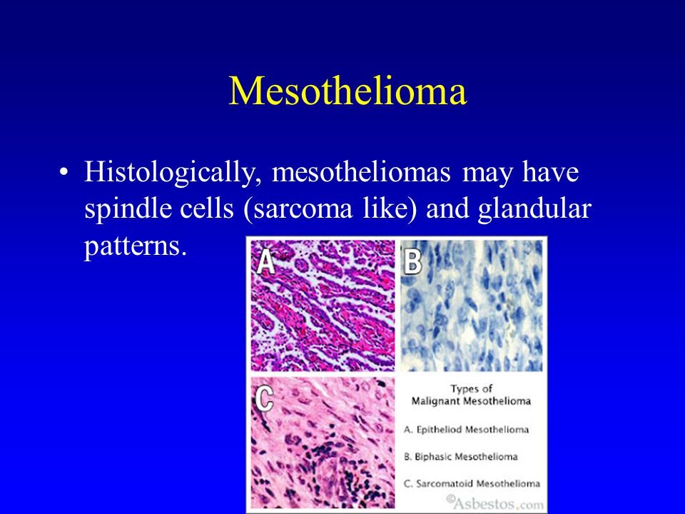
Pathologists perform tests, either histology or cytology, on samples. histology of the pleural, pericardial, and brain nodules showed sheets of tumour cells (fig 2), with areas of spindle cells and also epithelioid cells. The spindle cells stained strongly positively for cytokeratin. While the location of the cancer is important, the type of cells involved is significant as well. mesothelioma is difficult to diagnose by examining the macrostructure of the tumor. Pmc are more common than had previously been thought. The epithelioid form is occasionally confused with lung adenocarcinoma or metastatic carcinomas. histology is the study of tissues and cells under a microscope. This cell type produces aggressive tumors. Biphasic mesothelioma histology is specified as a mix of epithelial and also sarcomatoid cells. There are several variants of sarcomatoid mesothelioma, most notably the desmoplastic and lymphohistiocytoid subtypes. When physicians know the types of cells involved, they are able to personalize the treatment plan. It has a poor prognosis, with direct invasion of the chest wall or lungs and distant metastases.
Well differentiated papillary mesothelioma of the pleura (c38.4) 9052/3 epithelioid mesothelioma, malignant epithelioid mesothelioma, nos 9053/3 mesothelioma, biphasic, malignant. Signs and symptoms of mesothelioma may. Most of the lesion consists of a cellular spindle cell lesion with a collagenous appearance. It arises from the mesothelium. This cell type produces aggressive tumors.
Tumors Of The Lung And Upper Respiratory Tract Ppt Video Online Download from slideplayer.com mesothelioma is a type of cancer that develops from the thin layer of tissue that covers many of the internal organs (known as the mesothelium). Malignant mesothelioma is an uncommon malignant neoplasm that arises from mesothelial cells or multipotential subserosal mesenchymal cells of the pleura, peritoneum, pericardium, or tunica vaginalis of the testis. In situ hybridisation against p16 is a promising method of differentiating malignant mesothelioma from reactive mesothelium. Whichever cell type is a lot more dominant will certainly determine how the cells form and spread in the body. Pathologists perform tests, either histology or cytology, on samples. It arises from the mesothelium. Because of this distinct cell appearance, sarcomatoid mesothelioma is also known as "spindled mesothelioma" Biphasic mesothelioma, epithelioid mesothelioma, desmoplastic mesothelioma, sarcomatoid mesothelioma.
mesothelioma pathology is the study of tissue or fluid samples for the presence of mesothelioma.
Significant nuclear pleomorphism is relatively uncommon, more often seen in epithelioid tumors. It comes in three histological forms: histology is the study of tissues and cells under a microscope. Doctors look for mesothelioma biomarkers or diseased mesothelial cells. A, the spindle tumor cells were arranged in bundles and whirling patterns. Other names for this condition include sarcomatous mesothelioma, diffuse malignant fibrous mesothelioma, and spindle cell mesothelioma. mesothelioma is a cancer that forms in the mesothelium or the linings that surround various organs. This is where the nickname "spindled mesothelioma" In differentiating between sarcomatoid mesothelioma and a spectrum of spindle cell. Pleural mesothelioma starts in the lining inside the chest wall and around the lungs, peritoneal mesothelioma starts in the lining of the abdomen, pericardial mesothelioma starts in the lining membrane covering the heart, and. There are several variants of sarcomatoid mesothelioma, most notably the desmoplastic and lymphohistiocytoid subtypes. Basic concepts of pulmonary sarcomatoid carcinoma. Patterns of the epithelioid malignant mesothelioma include the deciduoid, clear cell, small cell, signet ring cell and pleomorphic cell patterns 4.
A solid pattern of mesothelioma on the right of this histologic section transitions to a spindle cell morphology on the left of the image in this predominantly sarcomatoid malignant mesothelioma mesothelioma histology. Its pathological characteristics include dense collagen fibers in a storiform pattern.


0 Comments